What is western blotting?
Overview of western blotting
Western blotting is the technique for identifying proteins of interest in a sample using gel electrophoresis to separate sample proteins. This technique has paved the way in biomedical science, providing a highly accurate approach to protein detection. Western blotting has been a critical tool in the detection of clinical signs of Lyme disease and HIV.
In this series we will break down the fundamentals of western blotting, providing an overview of equipment and techniques for running a successful blot. In this first piece, we will introduce you to the process of electrophoresis, and selecting your ideal transfer systems and buffer solutions.
What is a western blot?
Western blot is a process that detects specific proteins in a complex protein mixture extracted from cells or tissues. This biomedical procedure dates back to 1979 and is now the industry standard for protein analysis. Western blot has been used in breakthrough discoveries examining Lyme disease and Bovine Spongiform Encephalopathy (BSE), and complications with nutrient absorption.
There are three main steps in the workflow of western blotting: separation by size, transfer to stable support, and visualization using appropriate antibodies.
Simply put, western blotting is the process used to identify proteins via electrophoretic separation. This is done by transferring proteins from a gel onto a membrane where the proteins of interest can be accurately detected.
The membrane is treated with antibodies, binding epitopes onto the target protein. After a treatment with a second antibody, the targeted proteins in the sample can be visualized using detection techniques like the enhanced chemiluminescence (ECL) method.
It is somewhat of a lengthy process, requiring multiple incubation and wash steps. However, with a thorough understanding, you can save time, reagents, and samples with proper technique.
Electrophoretic separation of proteins
Electrophoresis is a process of separating particles within a gel substrate by influencing the charge of the particles with an electric field. This is a common process used by biochemists and molecular biologists to detect and isolate the presence of a specified protein in a complex sample mixture.
Electrophoresis is a technique used to separate proteins by charge. It can be used to investigate the composition of complex protein mixtures, or to verify the purity of protein samples. Electrophoresis separation can also serve to purify proteins for use in further applications.
For example, gel electrophoresis can be used for the separation of nanoparticles, and more specifically macromolecules (DNA, RNA, proteins). The gel is used as an anticonvective, or sieving medium during electrophoresis, which makes it easier to isolate and analyze macromolecules based on their size.
There are a few variables to consider when setting up an electrophoretic separation. Depending on your sample materials makeup and goals for your purified sample, it’s important to understand the correct parameters to run a successful separation. Variables such as pore size, protein charge, and shape determine the migration rate of the protein.
In polyacrylamide gel electrophoresis, proteins migrate in response to an electrical field through pores in a polyacrylamide gel matrix; pore size decreases with increasing acrylamide concentration. Gel electrophoresis offers an advantage, as the gel is easily poured and will not denature the sample. If handled properly, a sample can be recovered after processing. However, if the gel melts during electrophoresis, the buffer can become exhausted, causing sample materials to run in unpredictable forms.
Transferring proteins to a membrane
The next step in western blotting after electrophoresis is the transferring of proteins. Proteins will be moved from a gel matrix to a synthetic membrane support where it is bound, forming the blot. This is commonly executed via electrophoretic transfer.
The membrane and gel are placed together, with a barrier of filter paper placed between the two electrodes. Voltage is then applied between the electrodes, initiating the proteins to migrate to the membrane following the current of electricity.
The rate of elution of the proteins from the gel is determined by the strength of the electric field (measured in V/cm). The transfer conditions are reliant on the molecular weight of the target proteins and must be taken into consideration as to not under-transfer or over-transfer a sample. Based on your needs, there are a number of options available for transfer systems.
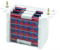
Tank Transfer Systems
Tank transfer is the most common method, offering the largest range of power settings and transfer times (30 minutes - overnight) and integrated cooling systems. These attributes make handling a broad range of molecule sizes possible. Gels and membranes are fully submerged in this system, requiring 1-12L of buffer and manual assembly.
Semi-Dry Systems
In the semi-dry systems, gels and membranes are placed between buffer-wetted filter papers that are in direct contact with flat-plate electrodes. Slightly easier to set up compared to tank systems, they require only some manual assembly. Due to the lack of cooling options, there are some limitations on power and transfer times (15-60 minute options). These systems are ideal for molecules within 30-120kD range and require only 250mL of buffer solution per blot.
Rapid-Transfer Systems
Rapid-transfer systems are the newest technology on the market offering the fastest transfer times (3-10 minutes). Similar to the traditional semi-dry system, rapid-transfer systems use filter papers and membranes that are pre-wet and prepared in single-use packaging.
This simplifies the assembly of the transfer stack and requires minimal to no manual setup. Semi-dry transfer techniques allow for more customization of transfer parameters, making them more practical for a broad range of sample molecular weights. No additional buffer reagent is required.
Blocking nonspecific sites
Next to washing and antibody incubation, blocking is the most critical phase of western blotting to guarantee accurate results. Blocking is key in the immunodetection phase of western blotting, as it prevents non-specific binding of antibody to the blotting membrane.
After the transfer of proteins from the gel, it’s important to block the remaining surface of the membrane to prevent nonspecific binding of the antibodies. This will ensure that results are readable.
Blocking is often made with a 5% Bovine Serum Albumin (BSA), or nonfat dried milk diluted in tris-buffered saline with Tween 20 (TBST) to reduce the background and make results clear. Depending on the detection labels being used, milk proteins are often not a compatible blocking solution. Understanding this compatibility will be important for a viable western blot.
Wash buffer formulations
Choosing the best wash buffer solution for blocking is necessary for a successful blot, as there are advantages and disadvantages to different solutions. This is decided by three factors: the protein of interest, the antibody, and the detection system being used.
Theoretically, you can use any buffer that does not have an affinity to bind with the target protein, or probe components. However, some buffer solutions will perform better than others. For example, if you use biotin and AP antibody labels along with antiphosphoprotein antibodies; BSA blocking solutions would be the ideal choice. Since milk contains casein (a phosphoprotein and biotin itself), a milk-based blocking solution would interfere with the assay results.
There are several common blocking buffers used in western blotting:
- Skim milk powder
- Bovine Serum Albumin (BSA)
- Normal serum (fetal serum from calf, rabbit, horse, or goat)
- Fish gelatin
- Purified casein
- Polyvinylpyrrolidone (PVP)
- Commercial buffers
The most common commercial buffer solution used is Sodium Dodecyl Sulfate (SDS), although SDS buffer presents some complications when working with some proteins. Proteins like Immobilon-P have been reported to pass through the plane of the membrane in the presence of SDS. In cases where the proteins have a tendency to precipitate, SDS works as a successful buffer.
When performing a western blot, some buffer solutions include a percentage of methanol, which will have benefits and limitations in some cases.
The methanol aids in stripping SDS from proteins transferred from denaturing SDS-containing polyacrylamide gels. This stabilizes the geometry of the gel during the transfer process and can increase the binding capacity of proteins to the Nitrocellulose (NC) within the membrane. Methanol also assists in the binding of proteins to the membrane. Methanol is not suited for transferring high molecular weight proteins or preserving enzyme activity. This is because methanol tends to shrink the gel.
Non-methanolic transfer is also ideal when transferring conformation sensitive antibodies. When working with a low-ionic strength buffer without methanol, it is important to incubate the gel prior to transfer for 30-60 minutes. By doing this, you avoid the risk of swelling that can distort the bands and make results difficult to read.
In most cases, it is good practice to incubate the primary antibody with BSA since it is typically needed in higher amounts than the secondary antibody. Doing this will allow the antibody to be reused if the blot does not provide a viable result.
Conclusion
Although Western blotting is a complex technique for protein detection, Avantor has the resources to make your western blotting project run smoothly. Avantor has you covered when it comes to selecting the best western blot technology on the market. We make sourcing simple, so you can focus on providing your team the ability to innovate with ease.